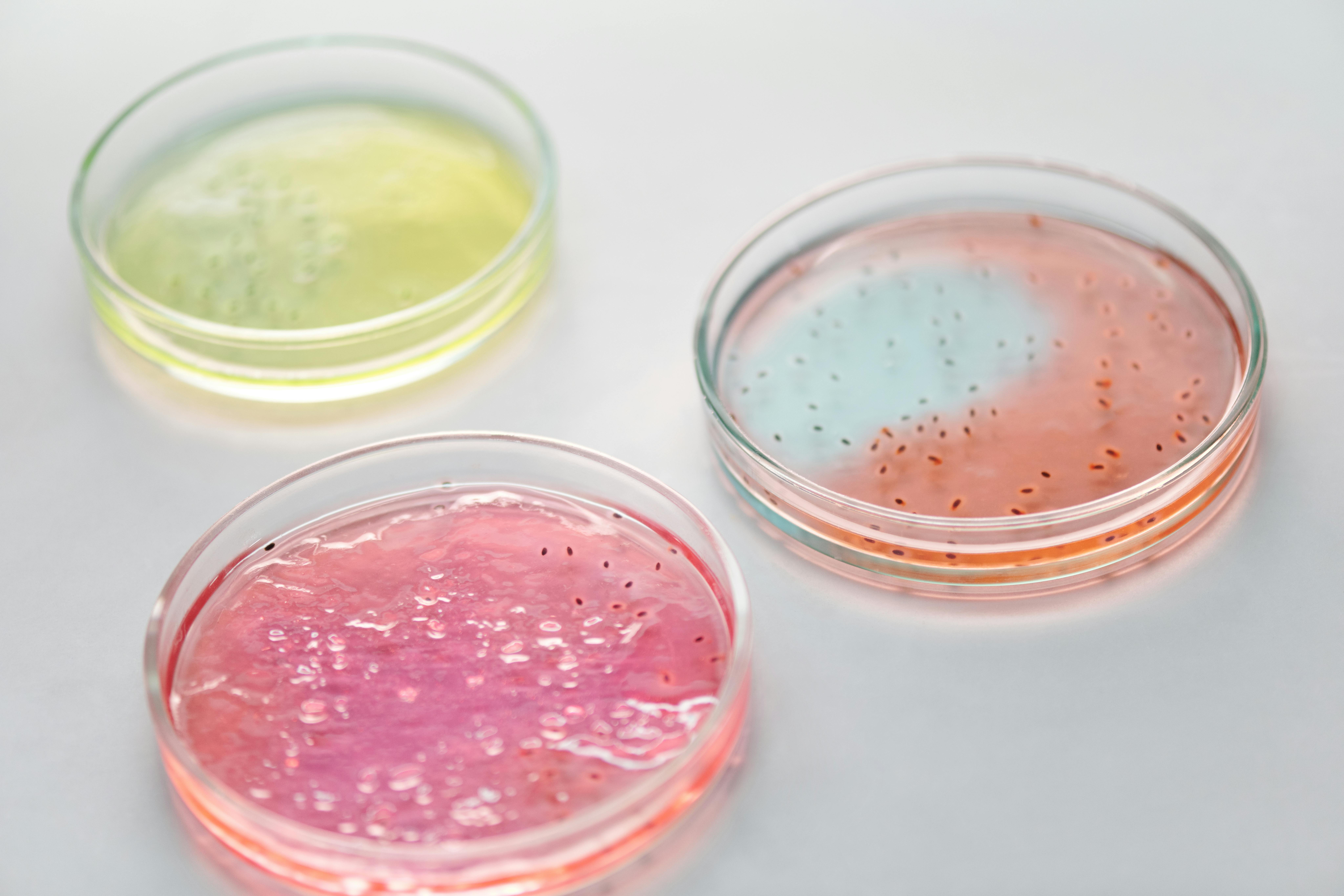
It lurks within us, a microscopic battleground within our own bodies. Our gut, a complex and vital organ, is constantly under siege from unwelcome invaders. Among the most formidable is Shigella, a cunning bacterium responsible for severe, often deadly, intestinal infections, especially in children. For years, scientists have struggled to truly understand how these tiny invaders wreak such havoc, largely because typical animal models just don’t quite capture the intricate ways these human-specific germs operate. But what if we could build a miniature, living replica of your gut, right in a lab dish, and finally watch the invasion unfold in real-time?
Breakthrough research published in Nature Genetics has done just that. Scientists have successfully used lab-grown mini-intestines, called organoids, to create the first comprehensive map of how Shigella bacteria take over human gut tissue. This isn’t just a fascinating scientific feat; it’s a game-changer for understanding and combating not only Shigella but potentially a whole host of other human-adapted pathogens that have been difficult to study. This gives us the detailed blueprints to the enemy’s battle plans, revealing its weak spots. The most startling discovery? Out of Shigella‘s roughly 5,000 genes, only about 100 are absolutely critical for it to establish a foothold and cause aggressive infection. This surprisingly short list of “must-have” genes is, as one of the study’s lead authors, Professor Mikael Sellin, put it, “a goldmine for understanding the progression of infections and for developing new treatments that can ‘turn off’ the bacteria’s pathogenic behavior.”
This is not a tale of a distant, abstract threat. Shigellosis, the disease caused by Shigella, is a very real danger, responsible for over 200,000 deaths annually, disproportionately affecting young children. The suffering it causes—severe inflammation, bloody diarrhea, and fever—can be debilitating. For too long, our ability to develop effective treatments has been hampered by the lack of realistic human models. Laboratory animals, while useful for many scientific endeavors, simply don’t mimic human physiology closely enough to fully understand how these bacteria operate within our bodies. This new research offers a powerful new way to view these microbial battles, opening doors to therapies that could, in the future, disarm these pathogens without resorting to broad-spectrum antibiotics that contribute to drug resistance.
Growing Mini-Guts to Fight Superbugs
At the core of this innovative study are “organoids”—tiny, three-dimensional structures that remarkably mimic the complexity of human organs. In this case, researchers grew intestinal organoids (from both the small and large intestine) using human stem cells. These stem cells were purified from surgical waste, ensuring the models accurately reflected actual human intestinal tissue. Unlike older methods using simpler lab-grown cells, these organoids maintain a more natural structure and function, providing a much more accurate battleground for studying bacterial infections.
To figure out which Shigella genes were essential for infection, the scientists used a clever technique called “transposon-directed insertion sequencing,” or TraDIS. Imagine having a massive library of Shigella bacteria, but in each “book,” one gene has been randomly “silenced” or deactivated. By infecting the human organoids with these specially engineered bacteria, the researchers could then see which “silenced” genes prevented Shigella from successfully invading the mini-guts. If a Shigella strain with a particular gene knocked out couldn’t infect the organoids, it strongly indicated that gene was crucial for its infectious capabilities.
A major challenge in such large-scale genetic investigations is dealing with “bottlenecks.” These occur in infection models when factors like the body’s defenses or competition for resources reduce the bacterial population, potentially causing the accidental loss of some bacterial mutants, regardless of their actual ability to cause disease. To overcome this, the research team refined their experimental procedures and developed a new statistical method called a “zero-inflated negative binomial (ZINB) distribution.” This advanced mathematical model helped them accurately account for the possibility of random bacterial loss, ensuring that their findings truly reflected a gene’s importance for infection, not just a fluke.
The main TraDIS screen involved 43 separate infection experiments, each using a “sub-library” of 2,000 to 3,000 unique Shigella mutants. After infecting the organoids for six hours, the bacteria that successfully invaded were recovered. The genetic makeup of these “survivors” was then compared to the original bacterial population. This comparison allowed the researchers to pinpoint genes that were either essential for colonization (meaning their absence made the bacteria less invasive) or, surprisingly, genes that actually hindered colonization (meaning their absence helped the bacteria).
Unmasking Shigella‘s Weaknesses
This rigorous approach yielded a comprehensive “genome-wide map” of Shigella genes vital for colonizing human intestinal tissue. They identified 105 genes on the Shigella chromosome and an additional 38 genes found on a special “virulence plasmid” (a small, circular piece of DNA often carrying genes important for infection). Many of these genes had never been linked to Shigella infection before, offering truly new insights.
Among the critical genes identified were those involved in the Shigella Type 3 Secretion System (T3SS), often described as the bacterium’s “molecular syringe.” This system is crucial for injecting bacterial proteins directly into human cells, a key step in infection. The study confirmed the importance of many known T3SS components, demonstrating the accuracy and depth of their screen. Beyond the T3SS, they found genes involved in stress regulation, various transporters, and even enzymes involved in sulfur metabolism.
Perhaps one of the most intriguing findings centered on two specific genes: mnmE and mnmG. These genes code for enzymes that modify transfer RNAs (tRNAs), which are essential molecules involved in creating proteins within the bacterium. When these mnmE and mnmG genes were disrupted, the Shigella mutants showed a significantly reduced ability to colonize the organoids—up to a tenfold decrease compared to normal Shigella. This was a crucial discovery because it pointed to a previously unrecognized way Shigella controls its ability to cause disease.
Further investigation showed that these mnmE and mnmG enzymes influence the production of many Shigella virulence proteins, especially those carried on the virulence plasmid. It turns out that genes important for Shigella‘s infectious power often use specific “codons”—three-letter sequences in DNA that act as instructions for building proteins—more frequently than other Shigella genes. The mnmE and mnmG enzymes ensure that the tRNAs can correctly “read” these specific codons, allowing the bacterium to produce the necessary virulence proteins efficiently. Without these modifications, the bacterium struggles to make its “weapons,” significantly weakening its ability to infect. This “post-transcriptional mechanism” (meaning control happens after the initial gene copying, at the protein-making stage) is a sophisticated way for Shigella to fine-tune its virulence.
Next Steps in Combating Infection
While this research marks a significant leap forward, it’s important to acknowledge its current limitations. The lab-grown organoids, while remarkably similar to human tissue, still lack certain components of a living human gut, such as nerves, blood vessels, and immune cells. The short infection window used in the screen (six hours) also means the study primarily focused on the initial stages of colonization. Processes related to long-term immune evasion or preventing cell death, which occur later in an infection, would require extended observation periods.
Despite these limitations, the authors emphasize that their work provides a “key proof-of-principle for functional genomics approaches in organotypic infection models.” This means the framework and techniques developed in this study can be built upon by future research, allowing scientists to investigate a wider range of pathogens and infection dynamics over longer timeframes. The insights gained here lay crucial groundwork for more complex investigations that could eventually lead to novel therapeutic strategies.
This research gives us a vital new weapon in our arsenal against infectious diseases. By understanding the precise genetic mechanisms Shigella uses to invade, we’re not just fighting an invisible enemy; we’re starting to learn its language, one gene at a time. This knowledge is not merely academic; it’s a direct path to developing more effective, targeted treatments that could save countless lives, particularly those of vulnerable children. It’s a powerful reminder that even the smallest breakthroughs in the lab can have the biggest impact on public health.
Paper Summary
Methodology Summary
The study utilized human intestinal organoids (miniature lab-grown gut tissues) derived from human stem cells purified from surgical waste material to model Shigella infection. To identify genes crucial for Shigella‘s ability to colonize human gut tissue, researchers employed transposon-directed insertion sequencing (TraDIS). This involved infecting organoids with a large library of Shigella mutants, each with a single gene randomly disrupted. A sophisticated Bayesian statistical model called Zero-Inflated Negative Binomial (ZINB) was developed to analyze the data, which helped account for experimental “bottlenecks” that could lead to misleading results. This allowed them to distinguish between genes genuinely important for infection and those whose absence was merely a result of random loss during the infection process. The findings were further validated using “barcoded consortium infections,” where mixtures of normal and mutant Shigella strains, each with a unique genetic tag, were used to infect the organoids, allowing for precise tracking of colonization efficiency.
Results Summary
The research identified 105 chromosomal genes and 38 virulence plasmid genes in Shigella that are essential for colonizing human intestinal epithelium. Many of these genes were previously unknown to be involved in Shigella infection. A particularly significant finding was the role of two tRNA-modification enzymes, MnmE and MnmG. Disruption of the genes encoding these enzymes severely reduced Shigella‘s ability to colonize the organoids (up to tenfold), indicating a novel mechanism of virulence control. Further investigation revealed that these enzymes are crucial for the efficient translation (protein production) of specific virulence proteins, especially those encoded on the virulence plasmid, which tend to use particular “codons” (genetic instructions) more frequently. The study demonstrated that MnmE/G-dependent tRNA modifications are essential for the accurate “reading” of these codons, thereby ensuring the production of the necessary proteins for Shigella‘s infectious behavior.
Limitations Summary
The study acknowledges that the human intestinal organoid model, while advanced, lacks some crucial components of a complete human gut, such as innervation (nerves), vasculature (blood vessels), and immune cells. Additionally, the relatively short infection window (six hours) primarily allowed for the study of early colonization events, meaning long-term infection dynamics, immune evasion, and cell death suppression mechanisms were not fully captured. These aspects would require further investigation with extended experimental setups.
Funding and Disclosures
The research was a collaborative effort of institutions including Uppsala University, Uppsala University Hospital, Helmholtz Institute for RNA-based Infection Research (HIRI), Toronto University, and Umeå University. Funding was provided by several grants from organizations such as ESCMID, the Carl Trygger Foundation, the Clas Groschinsky Memorial Foundation, the Bavarian State Ministry for Science and the Arts, the Swedish Research Council, the Swedish Foundation for Strategic Research, the SciLifeLab Fellows program, and Kempestiftelserna. The authors reported no competing interests.
Paper Publication Info
Article Title: A scalable gut epithelial organoid model reveals the genome-wide colonization landscape of a human-adapted pathogen Journal: Nature Genetics DOI: https://doi.org/10.1038/s41588-025-02218-x Received: 24 July 2024 Accepted: 1 May 2025 Published online: 12 June 2025


