Imaging has become so sophisticated that it can create pictures of human anatomy that seem to leap off the page. There’s still something, though, about dissecting the human body in three dimensions, and hands-on, that is worthy of awe. Medical students and physicians are beholden to donors for their forethought and generosity in willing their bodies for our study.
Consider the donors’ legacy. The four students assigned to each body use what we learn with the tens of thousands of patients for whom we care during our careers, and for the hundreds of students and residents whom we train, and their patients, and so on. I hope you enjoy learning a bit about the development of the human body and the beginnings of the plan for formation of some one-of-a-kind organs.
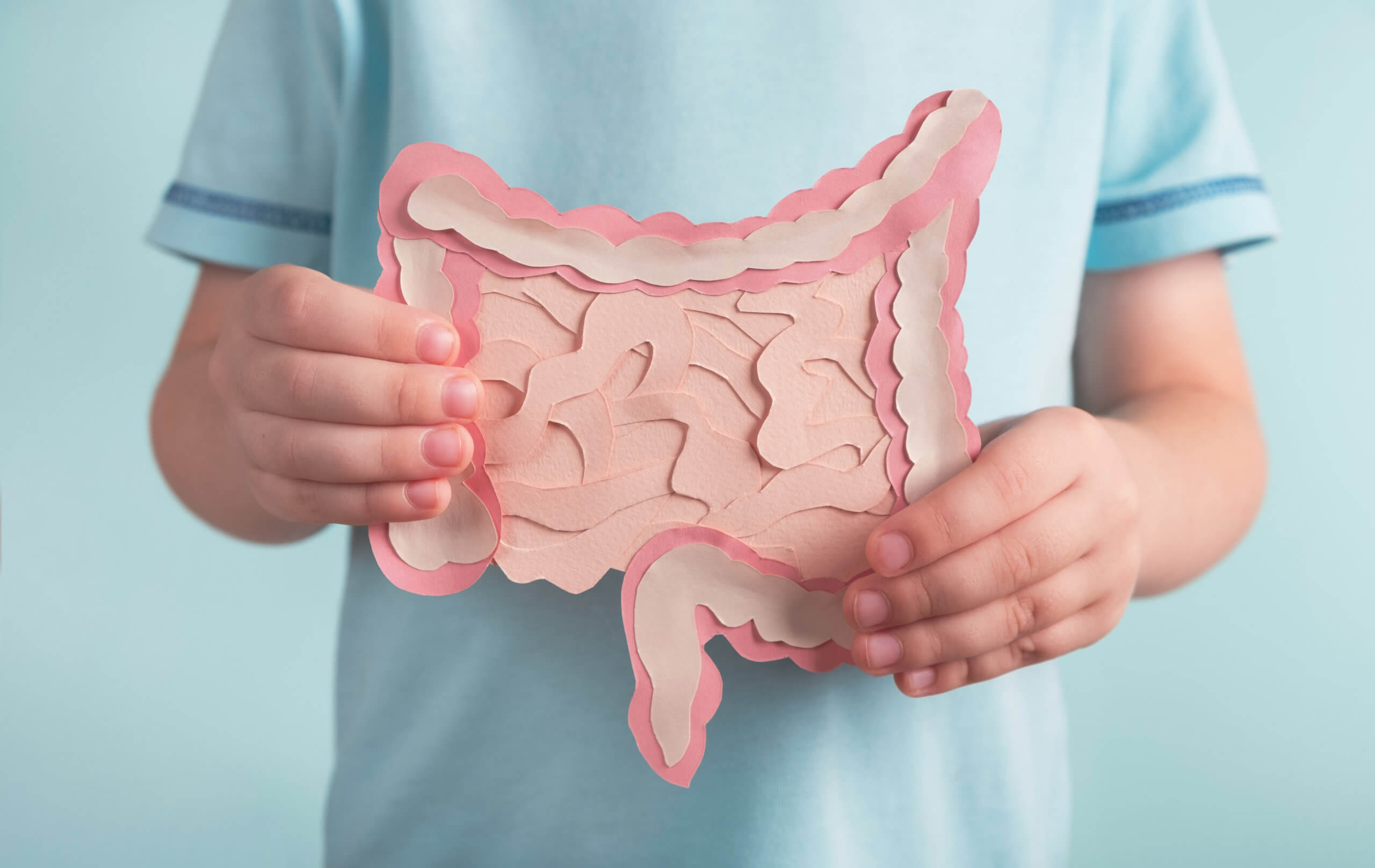
The gastrointestinal tract starts to differentiate while a child is still an embryo. It begins with a tube in the abdomen-to-be, rotating counterclockwise. Before now, what initiated that process was a mystery. A new study by scientists at Cornell University reveals, for the first time, that the gut rotation is initiated in two steps, by a transcription factor called Pitx2. A transcription factor is a protein that has a role in the reading and replication of genetic material, such as DNA and RNA. Further differentiation is triggered by mechanical forces within an elastic tissue that anchors the gut tube. This tissue later has a role in development of the blood and lymphatic vessels that supply the gut tube.
“The entire gastrointestinal tract is one tube that absorbs all of our nutrients, and it’s giant, so it has to loop to fit inside our body,” says Natasza Kurpios, associate professor of molecular medicine in the College of Veterinary Medicine at Cornell University, in a statement. “What we found years ago is that looping is highly conserved and it’s very, very regulated.”
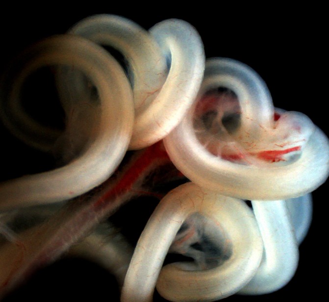
The researchers found that a type of sensor within the gut, TGF-beta, is dormant until it is activated by mechanical forces. In the gut, the mechanical force is generated by the dorsal mesentery, a structure which is attached to the gut tube and holds it in place. To perform the rotation, the tissue of the dorsal mesentery dramatically expands on what will be the child’s right side, and contracts on the left side. TGF-beta senses these forces, stimulating the looping of the gut tube.
There are other organs in the abdomen of which there is one of a type, such as the liver, gall bladder, stomach, pancreas, and spleen. The mechanisms and principles which apply to the development of the gut may lend insight into the development of other singular organs, with asymmetry of the left and right sides of the human body. One of the ways this knowledge can be used is to advance our understanding of birth defects and their management.
The study is published in the journal Science.